FIGURE 4.
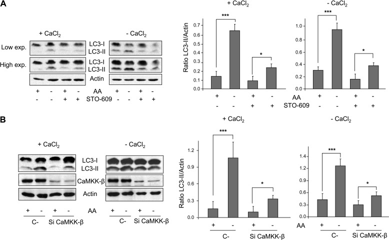
CaMKK-β is involved in the activation of autophagy by amino acid starvation. A, NIH3T3 cells were grown for 60 min in KH and AA with (+CaCl2) or without CaCl2 (in the last case, supplemented with 100 μm EGTA, −CaCl2) plus 100 μm leupeptin and 20 mm NH4Cl and in the presence or absence of 25 μm STO-609. In the last 30 min of incubation, AA were removed or not as indicated. Cell extracts (75 μg of protein) were analyzed by immunoblot using anti-LC3 (-I and -II) and, as a loading control, anti-actin antibodies. Low and high exposure (exp.) blots are shown. B, NIH3T3 cells were transfected with CaMKK-β (Si CaMKK-β) or with negative control (C-) siRNAs. After 72 h, cells were incubated as above and analyzed by immunoblot using the same antibodies as in A plus anti-CaMKK-β. Densitometric measurements of the LC3-II/actin ratios from three independent experiments in each case are also shown on the right of A and B. Differences were found to be statistically significant at p < 0.0005 (***) and p < 0.05 (*).

