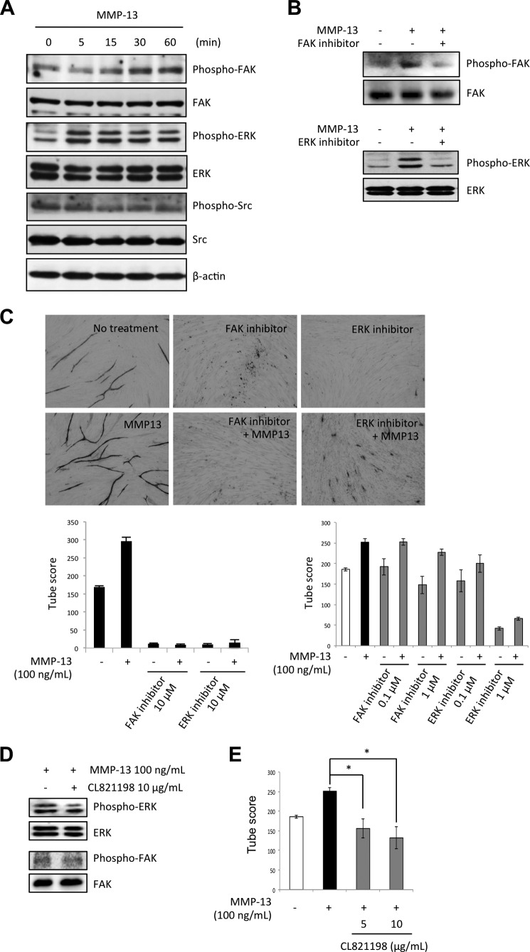
FIGURE 4.
MMP-13-promoted angiogenesis is mediated by FAK and ERK signaling pathway. A, levels of total and phosphorylated forms of FAK, Src, and ERK after treatment of HuhT1 cells with MMP-13 (100 ng/ml) shown by Western blotting. β-Actin expression was used as a loading control. HuhT1 cells were seeded on a culture dish. After incubation for 24 h, medium was changed to HuMedia without FBS and growth factors. After 4 h, the recombinant MMP13 protein (100 ng/ml) was added and the cells were incubated for indicated times. B, phosphorylated forms of FAK (Tyr-576/577), Src (Tyr-416) and ERK (Thr-202/Tyr-204) in the presence of MMP-13 (100 ng/ml) after treatment with 10 μm FAK inhibitor (FAK inhibitor 14) or 10 μm ERK inhibitor (U0126). Expression of total FAK or ERK was used as a loading control. C, upper panel shows the representative area of capillary tube formation by FAK inhibitor (FAK inhibitor, 14 or 10 μm) or ERK inhibitor (U0126, 10 μm) with or without MMP-13 (100 ng/ml) (×40). The lower left graph shows the average tubule score after 10 μm FAK inhibitor (FAK inhibitor 14) or 10 μm of ERK inhibitor (U0126) with or without 100 ng/ml of recombinant MMP-13 protein. The lower right graph shows the average tubule score after FAK inhibitor (0.1 and 1 μm) or ERK inhibitor (0.1 and 1 μm) with or without 100 ng/ml of recombinant MMP-13 protein. The values represent means of capillary tube score + S.D. based on three wells/data point in a single experiment. D, to examine the effect of protease inhibition on FAK and ERK phosphorylation, CL-821198, which is a selective inhibitor of MMP-13 through the binding to the S1′ pocket of MMP-13 with its morpholine ring adjacent to the catalytic zinc atom, was used. HuhT1 cells were seeded on a culture dish. After incubation for 24 h, medium was changed to HuMedia without FBS and growth factors. After 4 h, CL-821198 (10 μg/ml) and/or recombinant MMP13 protein (100 ng/ml) were added, and the cells were incubated for 1 h. Levels of total and phosphorylated forms of FAK and ERK was examined by Western blotting. E, capillary tube formation was examined by using an angiogenesis assay kit. HUVECs were treated with the recombinant MMP-13 protein with or without CL-821198 (5 and 10 μg/ml), and the medium was changed every 3 days. After 12 days, the cells were fixed and stained with anti-human CD31 antibody. The graph shows the average capillary tube score after treatment with recombinant MMP-13 protein. The capillary tube score was estimated with the Chalkley count method under a bright-field microscope. The values represent means of capillary tube score + S.D. based on three wells/data point in a single experiment. *, p < 0.05.

