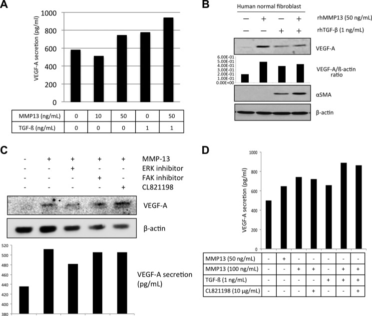
FIGURE 5.
VEGF-A secretion by MMP-13 treatment in fibroblasts. A, fibroblasts were seeded on a culture dish. After incubation for 24 h, medium was changed to DMEM without FBS. After 24 h, MMP-13 (0, 10, and 50 ng/ml) and TGF-β (1 ng/ml) with or without MMP-13 (50 ng/ml) were treated for 24 h. The concentration of VEGF-A in the culture medium was quantified with commercial ELISA kits according to the manufacturer's instructions. B, after treatment with MMP-13 (0, 10, and 50 ng/ml) or TGF-β (1 ng/ml) with or without MMP-13 (50 ng/ml) for 24 h, fibroblasts were collected. Expressions of VEGF-A, α-SMA, and β-actin were examined by immunoblotting. The densitometric analysis of VEGF-A expression was performed. VGEF-A/β-actin ratio is shown. C, HuhT1 cells were seeded on a culture dish. After incubation for 24 h, medium was changed to HuMedia without FBS and growth factors. After 4 h, the recombinant MMP13 protein (100 ng/ml) with or without 10 μm of FAK inhibitor (FAK inhibitor 14), 10 μm of ERK inhibitor (U0126) or 10 μg/ml of CL-821198 were added and the cells were incubated for 1 h. Expression of VEGF-A and β-actin were examined by immunoblotting. The concentration of VEGF-A in the culture medium was quantified with commercial ELISA kits according to the manufacturer's instructions. D, fibroblasts were seeded on a culture dish. After incubation for 24 h, medium was changed to DMEM without FBS. After 24 h, MMP-13 (0, 50, and 100 ng/ml) and TGF-β (1 ng/ml) with or without MMP-13 (100 ng/ml) were treated for 24 h. Moreover, we treated CL-821198 (10 μg/ml). The concentration of VEGF-A in the culture medium was quantified with commercial ELISA kits according to the manufacturer's instructions.

