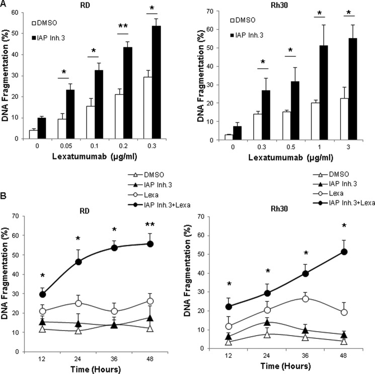
FIGURE 3.
IAP inhibitor 3 and lexatumumab cooperate to induce apoptosis. RD and Rh30 cells were treated with 1 μm (RD) or 2.5 μm (Rh30) IAP inhibitor 3 (IAP Inh.3) and/or the indicated concentrations of lexatumumab for 48 h (A) or with 0.2 μg/ml (RD) or 1 μg/ml (Rh30) lexatumumab (Lexa) and/or 1 μm (RD) or 2.5 μm (Rh30) IAP inhibitor 3 for the indicated time points (B). Apoptosis was determined by FACS analysis of DNA fragmentation of propidium iodide-stained nuclei. Means ± S.D. of three independent experiments performed in triplicate are shown. *, p < 0.05; **, p < 0.001 comparing cells treated with lexatumumab in the absence and presence of IAP inhibitor 3. DMSO, dimethyl sulfoxide.

