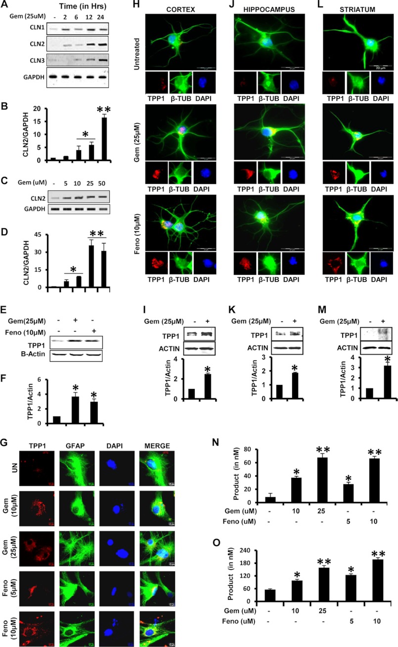
FIGURE 1.
Gemfibrozil and fenofibrate up-regulate TPP1 mRNA and functionally active protein in mouse brain cells. A and B, mouse primary astrocytes were treated with 25 μm gemfibrozil (Gem) in serum-free DMEM/F-12 for 2, 6, 12, and 24 h followed by monitoring the mRNA expression of Cln1, Cln2, and Cln3 by semi-quantitative RT-PCR (A) and qPCR (B) (for Cln2). C and D, mouse astrocytes were treated with different concentrations of gemfibrozil for 24 h under the same culture conditions followed by monitoring the mRNA expression of Cln2 by semi-quantitative RT-PCR (C) and real time PCR (D). E, mouse primary astrocytes were treated with 25 μm gemfibrozil and 10 μm fenofibrate (Feno) for 24 h under the same culture conditions followed by Western blot for TPP1. F, densitometric analysis of TPP1 expression (relative to β-actin) by gemfibrozil and fenofibrate treatment. G, mouse primary astrocytes were treated with different concentrations of gemfibrozil and fenofibrate under similar culture conditions and were double-labeled for TPP1 (red) and GFAP (green). UN, untreated. Scale bar, 10 μm. H, J, and L, mouse primary neurons were isolated from different parts of the brain and were treated with 25 μm gemfibrozil and 10 μm fenofibrate in neurobasal media containing B27-AO for 24 h and were double-labeled for TPP1 (red) and β-tubulin (β-TUB) (green) (H, cortical neurons; J, hippocampal neurons; L, striatal neurons.) DAPI was used to stain nuclei. Scale bar, 20 μm. I, K, and M, mouse neurons were treated with 25 μm gemfibrozil under same culture conditions for 24 h followed by Western blot for TPP1 (I, cortical neurons; K, hippocampal neurons; M, striatal neurons.) Graphs represent the densitometric analysis of TPP1 level (relative to β-actin). N, mouse primary neurons were treated with different concentrations of gemfibrozil and fenofibrate in B27-AO containing Neurobasal media for 24 h followed by the activity assay using cell extract containing 5 μg of total protein. O, mouse primary astrocytes were treated with different concentrations of gemfibrozil and fenofibrate in serum-free DMEM/F-12 medium for 24 h followed by the activity assay using cell extract containing 5 μg of total protein. All results are mean ± S.E. of at least three independent experiments. *, p < 0.05 versus control; **, p < 0.01 versus control.

