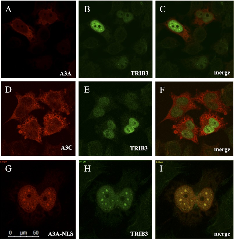
FIGURE 3.
Cell localization of the A3A and TRIB3. A–C, confocal microscopy yielding scant evidence of cytoplasmic and slightly nuclear A3A-V5 co-localizing with a nuclear 3×FLAG-TRIB3 in HeLa cells 36 h after transfection. D–F, A3C, TRIB3, and merge. G–I, addition of the SV40 NLS (residues PPKKKRKV) to the carboxyl terminus of A3A co-localization with TRIB3 is readily observed.

