FIGURE 2.
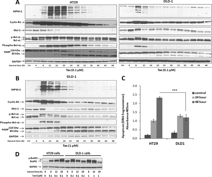
Death in mitosis is accompanied by phosphorylation of Bcl-xL and degradation of Mcl-1. A, HT29 (left panel) or DLD-1 (right panel) cells were released from single thymidine block, and after 4.5 h, treated with 0.1 μm Taxol (Tax) and harvested at the indicated time intervals. Immunoblots were performed with MPM-2 antibody or for the proteins indicated. GAPDH was used as a loading control. Phosphorylated Bcl-xL was detected by mobility shift (p-Bcl-xL) or reactivity with the phospho-Ser-62-specific antibody (phospho-Bcl-xL). Intact (116-kDa) and cleaved (85-kDa) species of PARP are shown. B, DLD-1 cells were released from single thymidine block, and after 4.5 h, treated with 1 μm Taxol and harvested at the indicated time intervals after treatment. C, HT29 or DLD-1 cells were untreated or treated with 0.1 μm Taxol for 24 or 48 h, and apoptosis was measured by cell death ELISA, as described under “Experimental Procedures.” Values represent mean ± S.D. (n = 3). *** = p < 0.001 (Student's t test). D, synchronized HT29 or DLD-1 cells were untreated or treated with either 0.1 μm or 1 μm Taxol for the times indicated (Harvest time (h)), and cell extracts were prepared and subjected to immunoblotting for BubR1 expression. BubR1 phosphorylation was detected by mobility shift (p-BubR1). GAPDH was used as a loading control.

