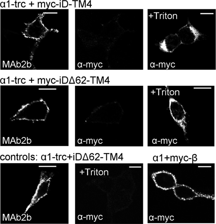
FIGURE 6.
Orientation of Myc-iD-TM4 and Myc-iDΔ62-TM4 within the plasma membrane. Confocal images of live stained cells are shown. HEK293 cells were transfected with either α1-trc + Myc-iD-TM4 or α1-trc + Myc-iDΔ62-TM4. The α1-trc was detected using MAb2b, and the Myc-iD-TM4 (Myc epitope is fused to the intracellular N terminus) was stained using a α-Myc antibody. As control, fixed and Triton X-100-treated cells showed intracellular protein (last panels in the top and middle rows). As negative control, cells transfected with α1-trc + iDΔ62-TM4 (without any tag) were stained with MAB2a for α1-trc and α-Myc for the iDΔ62-TM4 (last in permeabilized cells, +Triton X-100). Transfected cells with GlyRα1 + GlyRβ + gephyrin were used as positive control for the α-Myc antibody because the GlyRβ was tagged with a Myc epitope at the N-terminal extracellular part. Scale bars, 20 μm.

