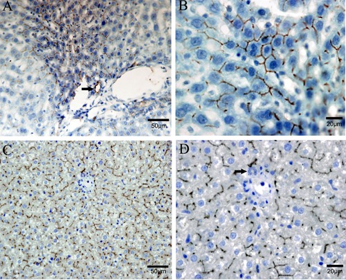
Figure 1.

Distribution of CM1 and CM2 antigens on rat liver tissue by immunohistochemistry analysis. A, B) Distribution of CM1 antigen on rat liver tissue. C, D) Distribution of CM2 antigen on rat liver tissue. Both CM1 and CM2 antigens were high positive on the hepatocyte canalicular membrane, but not on sinusoidal membrane or intracellular staining. Brown areas represented positive staining, blue areas represented hematoxylin counterstaining and arrow represented interlobular bile duct.
