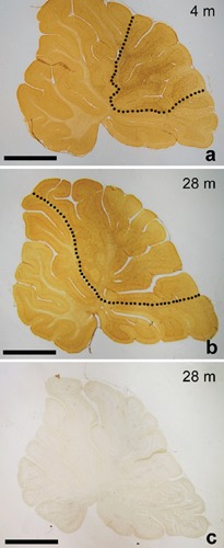
Figure 1.

a, b) Representative sections of the cerebellar vermis of 4- and 28-month-old rats showing the distribution of NG2 cells. The cerebellar area in which NG2 cells are uniformly and densely scattered (dotted line) is enlarged in aging animals; C) a section from a 28-month-old animal incubated with normal rabbit serum instead of the primary antibody. No immunoreaction is present. Scale bar: 2 mm.
