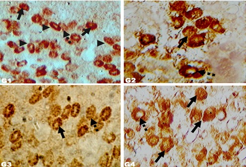
Figure 5.

Immunoreactive somatotrophs in molted layers. Immunohistochemical localization of GH cells (400 ×) in Zn induced molted layers of different groups: G1 (control, crude protein 16%; no other supplement); G2 (crude protein 18%; no other supplement); G3 (crude protein 16%; symbiotic at 85 mg L−1 drinking water); G4 (crude protein 16%; probiotic at 85 mg L−1 drinking water). Arrowheads: elliptical GH cells. Arrows: more densely stained immunoreactive GH cells. Dashed arrows: less densely stained immunoreactive GH cells.
