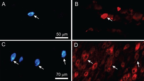
Figure 2.

Left: fluorescence photomicrographs of labeled nucleated neuronal profiles in superior cervical (A) and trigeminal (C) ganglia after fast blue deposition in supradiscal articular space of rat temporomandibular joint. Right: photomicrographs of same sections showing NPY (B) and CGRP (D) immunoreactive nucleated neuronal profiles after immunostaining with Cy3 conjugated streptavidin. Arrows indicate nucleated neuronal profiles retrogradely labelled with fast blue and displaying NPY-IR or CGRP-IR. Left and right photomicrographs sets are from identical sections viewed through separate filters. Scale bar = 50 µm (A and B) and 70 µm (C and D).
