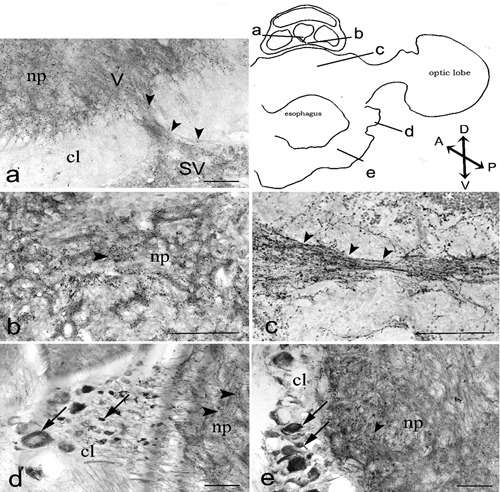
Figure 3.

cChAT immunoreactivity in the octopus supra and suboesophageal masses. The diagram of transverse section (obtained by camera lucida) show the locations of magnified photomicrograph: vertical lobe, V and subvertical lobe, SV (a); subvertical lobe (b); dorsal basal lobe (c), chromatophore lobe (d), pedal lobe (e). Arrows and arrowheads indicate IR cells and IR nerve fibers, respectively. Scale bars 100 µm. cl, cellular layer; np, neuropil. Axis: D, dorsal, V, ventral; A, anterior; P, posterior.
