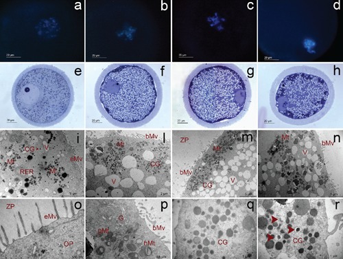
Figure 2.

Fluorescence (a, b, c, d), light (e, f, g, h) and transmission (i, l, m, n, o, p, q, r) micrographs representative of GV0 (a, e, i, o), GV1 (b, f, l, p), GV2 (c, g, m, q) and GV3 (d, h, n, r) oocytes. Mt, mitochondria; V, vacuoles; RER, rough endoplasmic reticulum; CG, cortical granules; eMV, erected microvilli; bMv, blent microvilli; ZP, zona pellucida; OP, ooplasm; pMt, pleomorphic mitochondria; hMt, hooded mitochondria; G, Golgi complex (from Lodde et al.41).
