Figure 4.
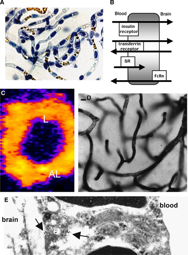
(A) Light micrograph of freshly isolated bovine brain capillaries stained with trypan blue, which stains nuclei blue and luminal red blood cells yellow. The capillaries are isolated free of adjoining brain tissue. (B) Schematic diagram of different brain endothelial receptors, including the insulin receptor, the transferrin (Tf) receptor, the scavenger receptor (SR), which mediates only the endocytosis from blood into the endothelial compartment for ligands such as acetylated low density lipoprotein; and the neonatal Fc receptor (FcRn), which mediates the asymmetric transcytosis of IgG molecules selectively from brain to blood, but not blood to brain. (C) Confocal microscopy of isolated rat brain capillaries showing the expression of the Tf receptor on both luminal (L) and abluminal (AL) membranes of the endothelium.100 (D) Light micrograph of rat brain removed after a 10-minute carotid artery infusion of a conjugate of 5 nm gold and the mouse OX26 monoclonal antibody (MAb) against the rat TfR. The staining of the capillary compartment represents TfRMAb present in the intraendothelial compartment of the brain.101 (E) Electron microscopy of rat brain after perfusion with the conjugate of gold and TfRMAb shows the MAb concentrated in intraendothelial vesicles (intracellular arrow), as well as MAb molecules undergoing exocytosis from the endothelial compartment to the brain interstitial space (extracellular arrow).101

