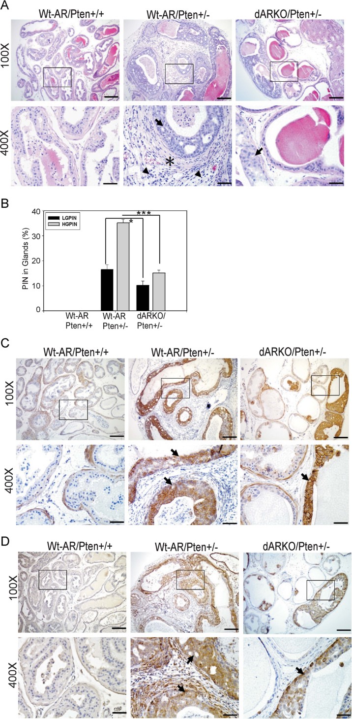
Figure 2. Loss of stromal fibromuscular AR leads to diminished PIN lesion development.
N = 6–7 mice per group
- Histological examination of 9-month-old Wt-AR/Pten+/+, Wt-AR/Pten+/− and dARKO/Pten+/− mouse DLPs subjected to H&E staining. The PIN lesions are presented as multi-layered epithelium and typical cribriform structures pointed-out by arrows. The asterisk marks the reactive stroma surrounding the PIN lesions and arrowheads indicate infiltrated immune cells located in the stromal compartments. Scale Bars = 200 µm (100×) and 50 µm (400×).
- Quantification of LG-/HG-PIN in Wt-AR/Pten+/+, Wt-AR/Pten+/− and dARKO/Pten+/− mouse DLPs at 9-month-old (N = 5 mice for each group). Data are presented as mean ± SEM. *p < 0.05, ***p < 0.001.
- IHC staining against p-Akt at Ser473. Arrows indicate p-Akt positive epithelium. Scale Bars = 200 µm (100×) and 50 µm (400×).
- IHC staining of p-S6RP (Ser235/236). Arrows indicate p-S6RP positive epithelium. Scale Bars = 200 µm (100×) and 50 µm (400×).

