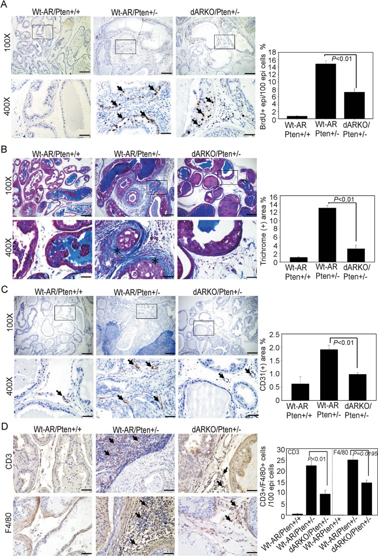
Figure 3. Stromal fibromuscular AR regulates epithelium proliferation, ECM remodelling, neovasculature formation and immune cell infiltration in Pten deficient mice.
N = 6–7 mice per group. Scale Bars = 200 µm (100×) and 50 µm (400×). The quantification results are in the right panels of each figure.
- BrdU staining was used to evaluate proliferative cells. Arrows indicate BrdU positive cells.
- Wt-AR/Pten+/+, Wt-AR/Pten+/− and dARKO/Pten+/− mouse DLPs were subjected to Masson's Trichrome staining to measure deposited collagen fibers that stain blue colors (asterisks).
- CD31 IHC staining was used to identify endothelial cells. Arrows indicate CD31+ endothelial cells.
- CD3 and F4/80 IHC staining of DLPs from three genotypes of mice were used to characterize infiltrated T-cells and macrophages, respectively. Arrows indicate CD3+ staining T-cells (upper panel) and F4/80+ macrophages (lower panel).

