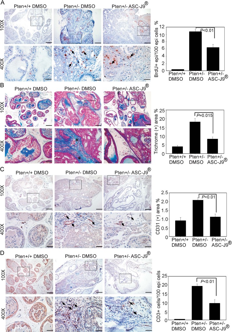
Figure 7. ASC-J9® impedes PIN lesion development and alleviates tumour-promoting microenvironment in Pten+/− mice.
N = 5–6 mice per group.
- The DLPs from three treated groups of mice were subjected to histological characterization by BrdU staining (left panel) and the quantification results are provided in the right panel.
- Pten+/+ DMSO, Pten+/− DMSO and Pten+/− ASC-J9® treated mouse DLPs were subjected to Masson's Trichrome staining to measure deposited collagen fibers. The blue colors indicate collagen fibers defined by asterisks and quantification results are shown in the right panel.
- CD31 IHC staining. Arrows indicate CD31+ endothelial cells in the stromal compartment and quantification results are shown in the right panel.
- CD3 IHC staining. Arrows indicate CD3+ T-cells and quantification results are shown in the right panel.

