Fig. 2.
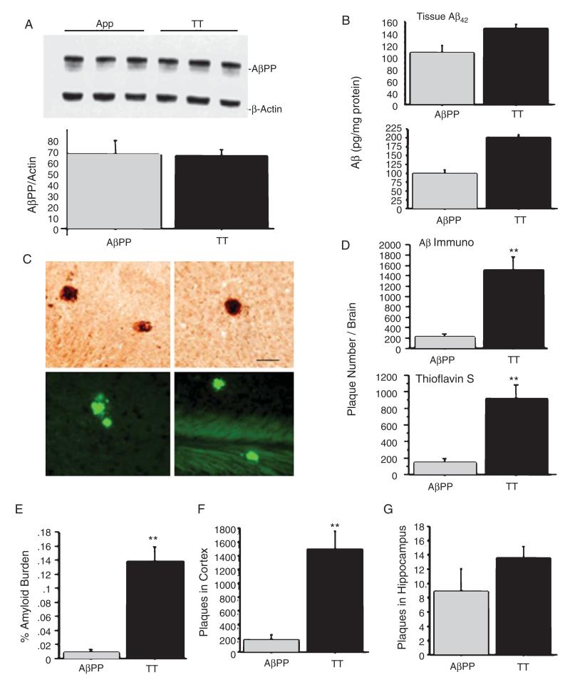
Analysis of AβPP and Aβ deposition in TT and AβPP+ mice. A) AβPP levels were similar in TT and AβPP+ mice (an unpaired two way Student-Test, p = 0.93) at 6 months of age. B) ELISA analysis shows that TT mice had higher levels of both soluble (p = 0.033) (upper panel) and insoluble (p = 0.0024) (lower panel) Aβ42 levels at 6 months of age compared to levels in AβPP+ mice. C) Aβ immunohistochemical staining revealed substantial Aβ plaque deposition in the cortex of TT mice at 6 months of age (upper panels, Bar = 50 μm), but not at 3 months of age. Thioflavine staining revealed a similar distribution of plaque deposition (lower panels). D, E) Quantitative analysis of Aβ plaque deposition indicates a significant effect of genotype on Aβ plaque number ((p < 0.0001) by immunostaining and thioflavine S staining, D) and amyloid burden (p < 0.0001, E). F, G) TT mice showed dramatic increases in plaque numbers and burden in the cortex, but not in the hippocampus, as compared to AβPP+ mice (**p < 0.01).

