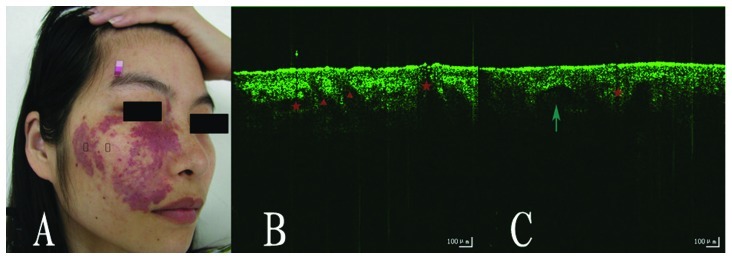
Figure 3.
(A) Image of a patient with PWS of the proliferative type. OCT image of (B) normal skin and (C) the PWS lesion. The test sites are marked by rectangles, the vessel is marked by an arrow, the sebaceous glands and hair follicles are marked by a triangle and star, respectively. PWS, port wine stain; OCT, optical coherence tomography.

