Fig. 5.
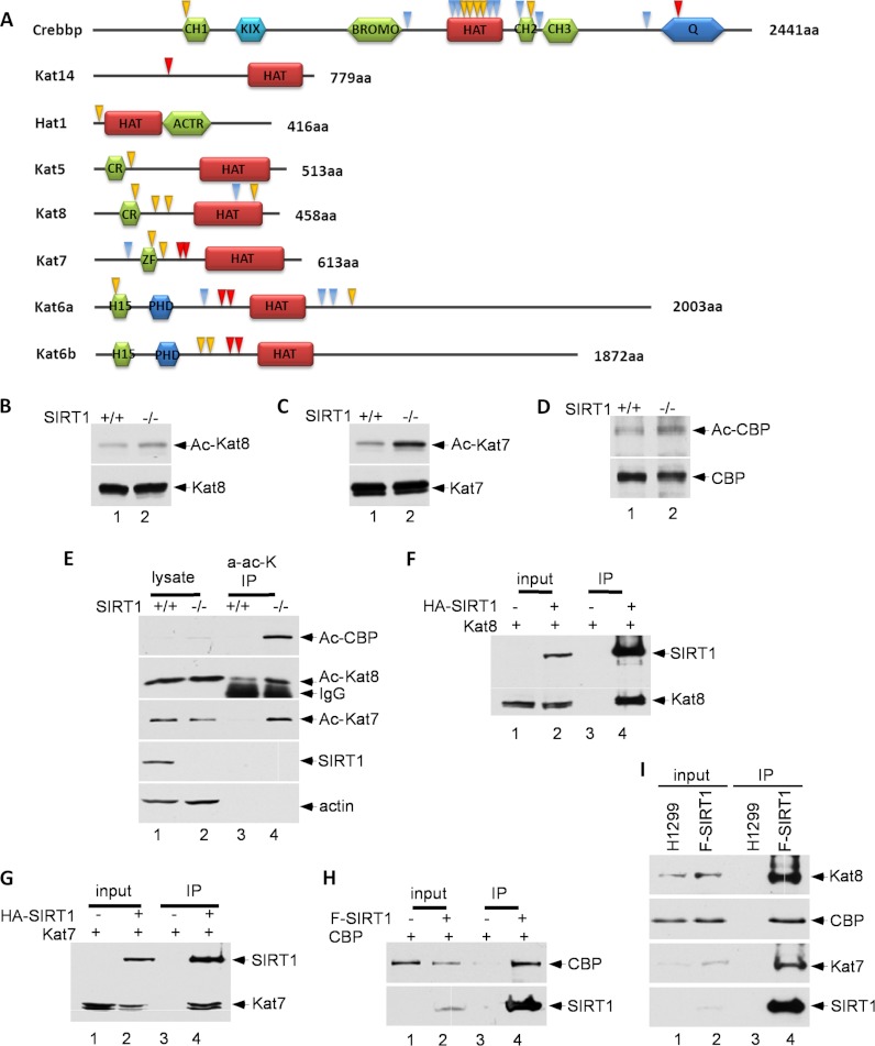
SIRT1 deacetylates histone acetyltransferases (HATs). A, Schematic representation of HATs. The acetylated lysine residues identified through SILAC are indicated by arrows. Yellow arrows indicate a site quantification ratio (SIRT1 KO/WT) greater than 2, red arrows indicate sites only identified in SIRT1 knockout cells, and blue arrows indicate all other sites. B, C, and D, MEF cells (SIRT1 KO and WT) were treated with 1 μm trichostatin A (TSA) for 16 h to prevent the interferences from class I and II HDACs. The protein lysate containing 1 μm TSA and 10 mm nicotinamide was immunoprecipitated using corresponding antibodies, and immunoblotted with an anti-acetyllysine antibody. E, MEF cells were treated with 1 μm TSA for 16 h. The protein lysate was immunoprecipitated using anti-acetyllysine antibody and immunoblotted with antibodies of interest. F, G, and H, Protein extract from whole transfected 293 cells was immunoprecipitated and immunoblotted with antibodies of interest. I, Protein extracts from whole Flag-SIRT1/H1299 cells and H1299 parental cells were immunoprecipitated with M2 beads and immunoblotted.

