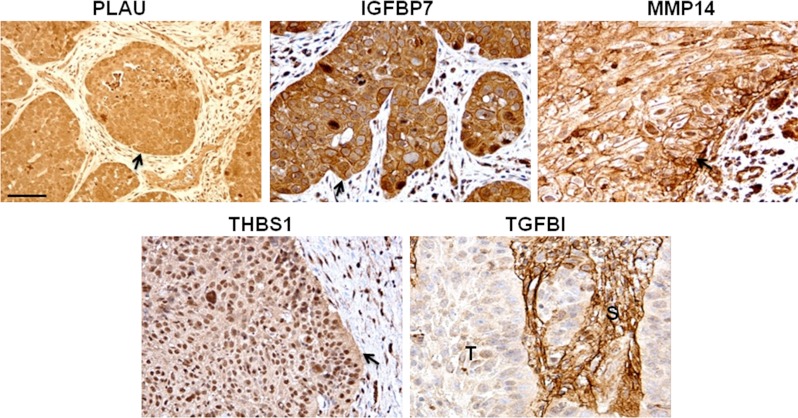
Fig. 4.
Verification of selected markers in HNSCCs by immunohistochemistry. Representative images shown. Scale bar 50 μm. Arrows and (T) indicate tumor tissue; (S) indicates stroma. All markers except TGFBI were localized in the cytoplasm of tumor cells and had some degree of expression at the plasma membrane. In contrast to the other four markers, TGFBI was predominantly expressed in stromal cells and had no or weak expression in the majority of tumor cells.

