Fig. 3.
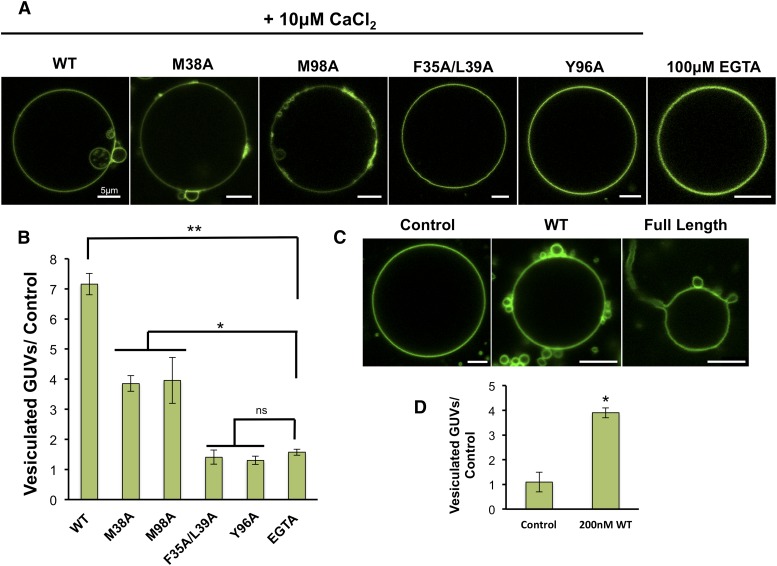
The GUV assay demonstrates the C2 domain's ability to induce membrane budding. A: Experiments with POPC:POPE:POPS (60:20:20) GUVs were performed in triplicate with a minimum of 60 GUVs assessed per measurement to provide a quantitative representation of membrane curvature changes shown in B. B: Quantitative representation of GUV vesiculation induced by C2 domain and respective mutants. WT induced significant vesiculation of GUVs in the presence of 10 μM CaCl2 compared with control experiments performed in 100 μM EGTA. M38A and M98A displayed some induction of GUV vesiculation and membrane reorganization but to a significantly lesser extent than WT. F35A/L39A and Y96A did not appreciably induce GUV vesiculation when compared with the control. C: The WT C2 domain and full-length cPLA2α were assessed at 200 nM protein concentration for their ability to induce membrane curvature changes in the presence of 500 nM CaCl2 to GUVs containing POPC:POPE:POPS (60:20:20). The WT C2 domain induced substantial vesiculation, whereas full-length cPLA2α induced vesiculation and long tubule formation from GUVs. D: Quantification of vesiculation in control versus WT C2 experiments shown in C. The P value for each protein was determined in comparison to the control in C and D (ns, not significant; *P < 0.001; **P < 0.0001) using an unpaired Student t-test. Scale bars = 5 μm.

