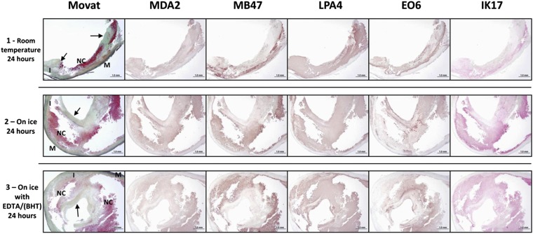
Fig. 10.
Immunostaining of oxidation-specific epitopes represented by antibodies MDA2, MB47, LPA4, EO6, and IK17 of fresh human carotid endarterectomy specimens. Prior to paraffin embedding, the endarterectomy specimens were manually cut into three sections (proximal, mid, distal) because the specimens were not symmetrical, and due to some fragmentation during removal, the immunostained sections are of different size. Specimens were then handled according to the following procedures: (Row 1) Proximal specimen stored at room temperature for 24 h in PBS (phosphate buffered saline. Staining for MDA2 is weakly present in predominantly the necrotic core. MB47 staining is more intense and seen in similar areas as for MDA2. Expression of LPA4 and EO6 is located in the extracellular matrix regions of the fibrous cap and the necrotic core. IK17 is positive in the late necrotic core. Fibrous cap (arrows) adjacent and partially overlying a late necrotic core (NC). Movat pentachrome; I, intima; M, media. (Row 2) Mid specimen stored on ice for 24 h in PBS. MDA2 expression is present within the necrotic core. MB47 expression is more intense and is located within the areas as seen in the MDA2 staining. The localization of LPA4 and EO6 expression is similar as described for row 1, mostly in the extracellular matrix regions of the fibrous cap and the necrotic core. IK17 is strongly positive in the large late necrotic core. (Row 3) Distal specimen stored on ice in EDTA/BHT antioxidant for 24 h. MDA2 is weakly present in the necrotic core, whereas MB47 expression is more intense. Expression of LPA4 and EO6 is mostly seen in the extracellular matrix regions of the fibrous cap and the necrotic core, and the intensity of the staining is comparable to the staining in row 2. IK17 is strongly present in the late necrotic core. MDA2, MB47, LPA4, EO6: Immunoperoxidase (brown reaction product). IK17: Alkaline phosphatase (red reaction product). Magnification ×100.

