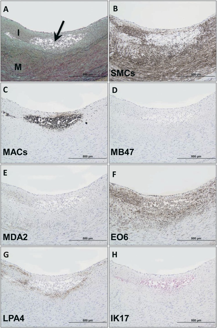
Fig. 2.
Immunostaining of OSE in a human coronary intimal xanthoma. A: Intimal xanthoma recognized as an early superficial lesion rich in macrophages without evidence of necrosis. Arrow, Movat pentachrome; I, intima; M, media) B: Areas of SMC (anti-SMC α-actin). C: Localization of CD68 staining for macrophages, which do not overlay regions of SMCs. D, E: Relative absence of staining for apoB-100 (MB47) and MDA epitopes (MDA2), respectively. F: Strongly positive OxPL (E06) staining colocalizes with macrophages and nearby SMCs of the neointima. G, H: apo(a) (LPA4) and IK17 epitopes, respectively, are expressed in select populations of macrophages. B–G: Immunoperoxidase (brown reaction product). H: Alkaline phosphatase (red reaction product). Magnification ×200.

