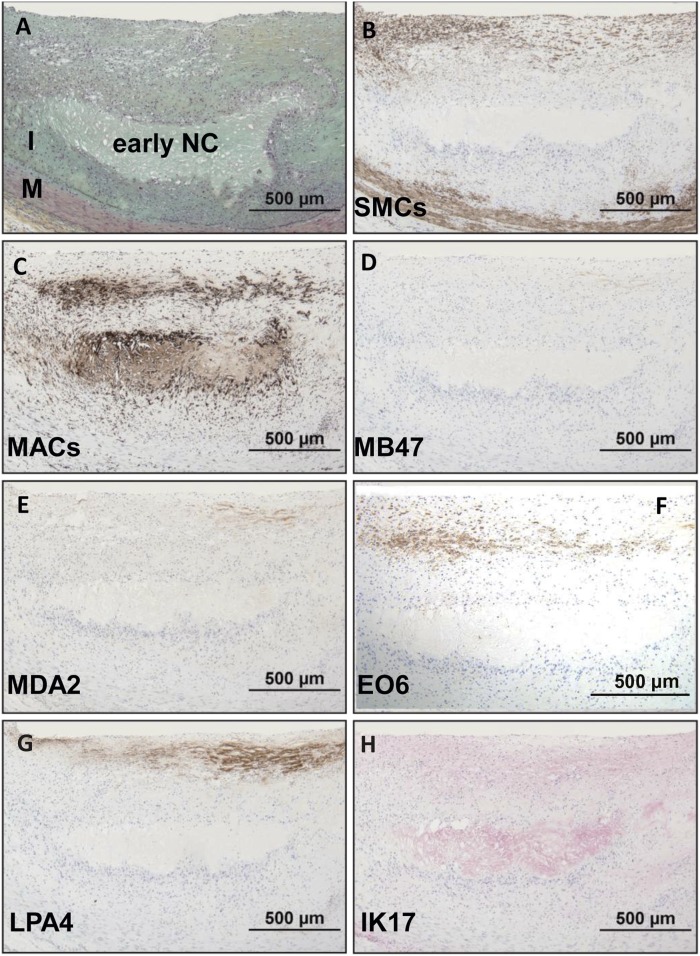
Fig. 4.
Immunostaining of OSE in a human coronary lesion recognized as early fibroatheroma. A: Human coronary lesion shows a superficial plaque with an early necrotic core (NC) characterized by macrophages, free cholesterol, and extracellular matrix. Movat pentachrome; I, intima; M, media. B: α-Actin-positive SMCs are mostly localized to the medial wall and fibrous cap. C: Localization of CD68 staining shows positive intense macrophage staining of the fibrous cap and necrotic core. D, E: Negative staining for MD47 and MDA2, respectively. F: E06 is found primarily localized to macrophages of the fibrous cap. G: Expression of LPA4 is primarily localized to the extracellular matrix of the fibrous cap. H: Weak to moderate staining of IK17 is seen in the necrotic core. B–G: Immunoperoxidase (brown reaction product). H: Alkaline phosphatase (red reaction product). Magnification ×200.

