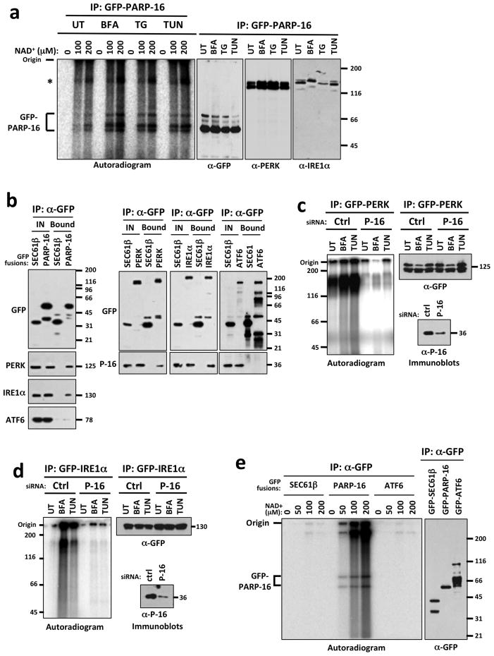
Figure 3. PERK and IRE1α are (ADP-ribosyl)ated in a PARP16-dependent manner during the UPR.
MW (kD) at right of blots. (UT)= untreated; (BFA)= Brefeldin A treated; (TG)= Thapsagargin treated; (TUN)= Tunicamycin treated. a, Autoradiogram of EMAA showing ADP-ribose incorporation. Immunoblots of GFP-PARP16 precipitates are shown at right. Asterisk= high MW NAD+ incorporation. n = 5; 0.01 < p of fold increase < 0.05 for all stressors. b, ER microsome based co-immunoprecipitation assays of GFP-fusion proteins. Shown are immunoblots of precipitated GFP fusions. c–d, EMAA using control or PARP16 knock-downs. Shown are autoradiogram and immunoblots of GFP-PERK (c) or GFP-IRE1α immunoprecipitates (d). For both (c) and (d), n=4; 0.005 < p of fold increase < 0.05 for all stressors. PARP16 immunoblots of control and PARP16 knock-down lysates are shown. e, EMAA for SEC61β, ATF6 and PARP16. Shown are autoradiogram and immunoblot of the immunoprecipitated GFP fusions. n=2.

