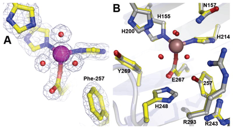
Figure 2.

Structure of the Y257F variant in the resting state (A) and comparison of the active site environments (B) for the Y257F variant (PDB 4GHC) with that of FeHPCD (PDB 3OJT). The blue 2Fobs Fcalc electron density map is contoured at 1.6 σ. Atom color code: gray, carbon (FeHPCD); yellow, carbon (Y257F); blue, nitrogen; red, oxygen (Y257F); dark red, oxygen (FeHPCD); purple, iron (Y257F); bronze, iron (FeHPCD). Cartoons depict secondary structure elements for FeHPCD (gray) and Y257F variant (light blue).
