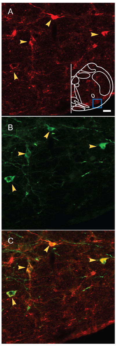
Figure 1. AAV2 viral targeting of preBötC Sst neurons.

Double immunostaining of Sst and EGFP shows the colocalization of both markers in preBötC. A: Sst-ir neurons. Blue box in outline represents area where images were acquired. B: EGFP-ir neurons. C: Colocalization of both markers indicated by yellow arrows. Magenta-green copy of this figure is available as Supporting Figure 1. Scale bar = 20 μm. [Color figure can be viewed in the online issue, which is available at www.interscience.wiley.com.]
