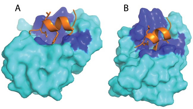
Figure 1.
Molecular model of the human p53 Mdm2 complex (A) and predicted placozoan p53. The Mdm2 complex (B). p53 is shown as orange ribbon/sticks, and Mdm2 is shown in cyan; the dark blue regions are conserved Mdm2 residues in contact with p53. Reproduced from Research Highlights: Protein’s billion-year history. Nature. 2010;463(7280):404.

