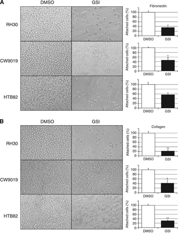
Figure 1.
Cell adhesion to fibronectin- or collagen-coated plates. Images showing a representative field of attached cells for each cell line in the presence (GSI) or absence (DMSO) of γ-secretase inhibitor and plots representing the quantification of cells attached to fibronectin- (A) or collagen-coated (B) plates. All samples were evaluated in triplicate. Student’s t-test significance: *P<0.05, **P<0.005.

