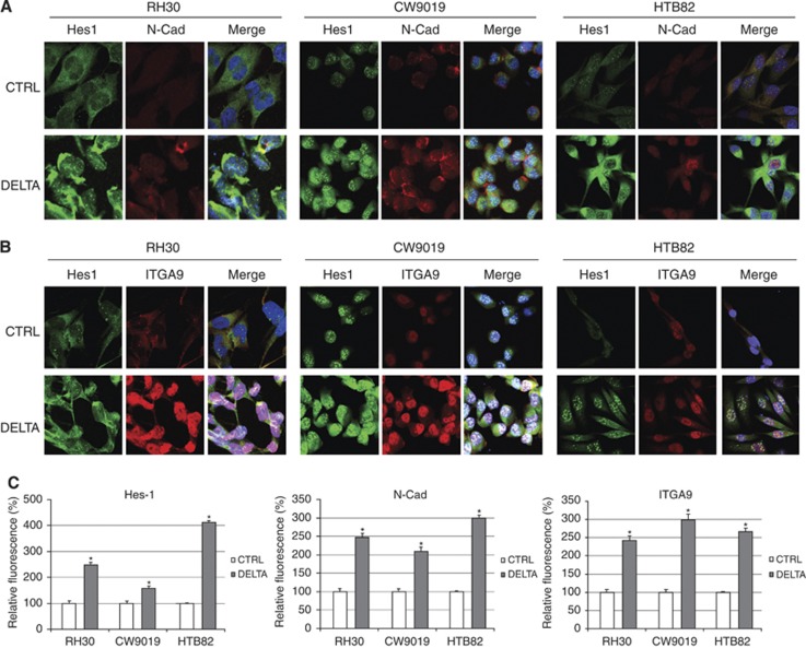
Figure 4.
N-cadherin and α9-integrin immunocytochemistry. Immunocytochemistry with anti-Hes1 and N-cadherin (N-Cad) antibodies (A) or Hes1 and α9-integrin (ITGA9) antibodies (B) in cells transfected with Delta or control vector (CTRL). Staining was as follows: Hes1 (green), N-cadherin and α9-integrin (red) and nuclei (blue). (C) Relative fluorescence quantification for Hes1, N-cadherin and α9-integrin. Student’s t-test significance: *P<0.05.

