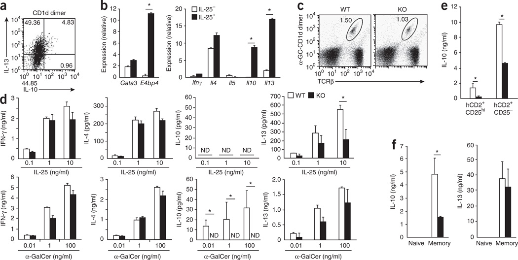
Figure 6.
Impaired production of IL-10 and IL-13 by CD4+ T cell subsets lacking E4BP4. (a) ICS detection of IL-13- and IL-10-producing cells among whole BALB/c splenocytes stimulated for 48 h with the α-GalCer–CD1d dimer (CD1d dimer). (b) Quantitative RT-PCR analysis of gene expression among total RNA from IL-17RB+ NKT cells generated from IL-17RB+α-GalCer–CD1d+TCRβ+ cells sorted from spleen cells and cultured for 24 h in the presence (IL-25+) or absence (IL-25−) of IL-25 together with bone marrow–derived DCs induced by granulocyte-macrophage colony-stimulating factor. (c) Flow cytometry analysis of NKT cells from the spleens of E4bp4−/− mice and their E4bp4+/+ littermates. Numbers adjacent to outlined areas indicate percent α-GalCer–CD1d+TCRβ+ cells. (d) ELISA of cytokine production in IL-17RB+ NKT cells derived from E4bp4−/− mice and their E4bp4+/+ littermates (C57BL/6 background) and stimulated for 24 h with various concentrations of IL-25 (top) or α-GalCer (bottom) in the presence of bone marrow–derived DCs induced by granulocyte-macrophage colony-stimulating factor. ND, not detected. (e,f) ELISA of cytokines in culture supernatants of Foxp3+CD4+ (CD25− or CD25hi) T cells (e) and naive (Foxp3−CD62LhiCD44lo) and memory (Foxp3−CD62LloCD44hi) CD4+ T cells (f) purified from reporter mice expressing Foxp3 from human CD2 (hCD2), on an E4bp4+/+ or E4bp4−/− background, then stimulated for 48 h with mAb to TCRβ and mAb to CD28. *P < 0.01 (Student’s t-test). Data are representative of two experiments (a) or three experiments (b–f; mean and s.e.m.).

