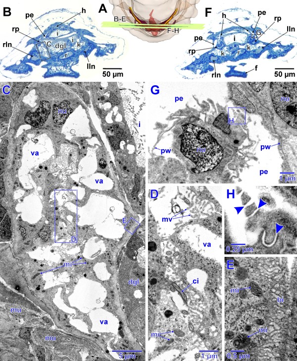
Figure 8.
Advanced stage of metanephridial system development in Lepidochitona corrugata (spm 8). A. Posterior part of specimen from dorsal with transparent external surface and planes (ortho slices) of section images shown in B–H. B. Total LM cross section through the anlagen of the metanephridial system with stippled rectangles indicating the position of details given in the TEM sections C–E. C. Right metanephridial kidney. Stippled rectangles mark areas shown in D and E. D. Metanephridial lumen. E. Surface of kidney with basal infoldings. F. Total LM cross section through the anlagen of the metanephridial system with stippled rectangles indicating the position of details given in the TEM sections G and H. G. Ultrafiltration site in the atrial wall. Stippled rectangle marks area shown in H. H. Ultrafiltration site, arrowheads indicating ultrafiltration slits. bi, basal infoldings; ci, cilium; dgl, digestive gland; f, foot; h, heart; i, intestine; k, kidney; lln, left lateral nerve cord; mi, mitochondria; mu, muscle fibers; mv, microvilli; nu, nucleus; pe, pericardium; pw, pericardial wall; r, rectum; rln, right lateral nerve cord; rp, renopericardial duct; va, vacuole.

