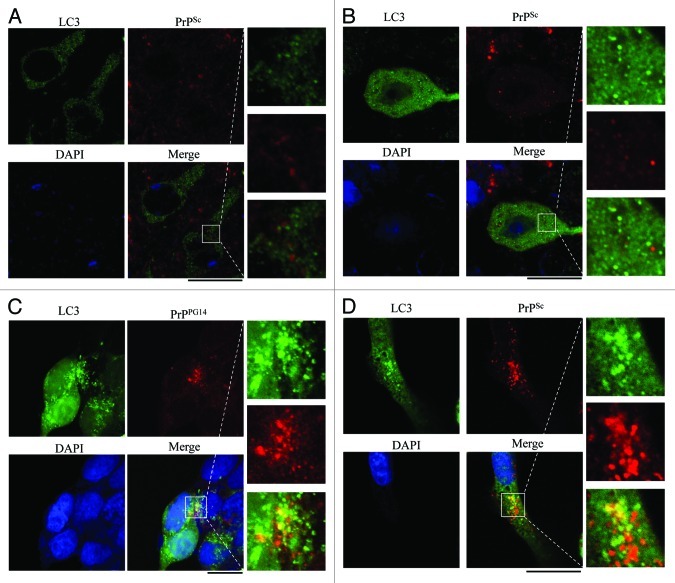
Figure 11. Assays of the colocalization between autophagosome and PrPSc in the brain sections of 263K-infected hamsters at terminal stage and in the prion cell models. The paraffin sections of scrapie 263K-infected hamster’s brain [(A) cortex, (B) the cerebellum] were double-stained immunofluorescently for LC3 and PrPSc. (C) HEK293 cells transiently cotransfected with pcDNA-PrP-PG14 and pEGFP-LC3 were stained immunofluorescently for PrP after treated with 400 nM bafilomycin A1. (D) SMB-S15 cells transfected with pEGFP-LC3 were stained for PrPSc after treated with 400 nM bafilomycin A1. The magnification views were shown in the right of each picture. The images of LC3 (green), PrPSc or PrPPG14 (red), DAPI (blue) and merge are indicated above. Scale bar: 20 μm.

An official website of the United States government
Here's how you know
Official websites use .gov
A
.gov website belongs to an official
government organization in the United States.
Secure .gov websites use HTTPS
A lock (
) or https:// means you've safely
connected to the .gov website. Share sensitive
information only on official, secure websites.
