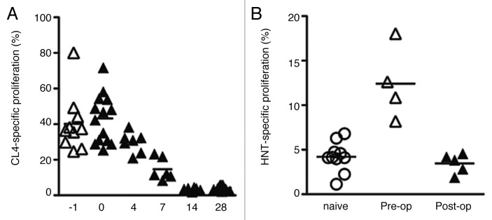
Figure 3. HA-specific CD8+ and CD4+ T-cell proliferation in the draining nodes pre- and post-complete AB1HA tumor resection. (A) CL4 CD8+ T cells were transferred at day 16 of tumor growth (day -1), at the time of surgery (day 0), or on post-operative days: 4,7,14, and 28. Each data points depicts proportion of proliferating CFSE+ CL4 T cells detected in pooled inguinal and axillary DLN from an individual mouse. Mean proliferation of group indicated by bars. (B) HNT proliferation was assayed in BALB/c with established (day 16) AB1HA tumors, and two weeks after surgery. Experiment was performed once, with five animals in the pre-operative and 2 week post-operative groups (n = 10 for naïve group). Each data points depicts proportion of proliferating CFSE+ HNT T cells detected in pooled inguinal and axillary DLN from an individual mouse.

An official website of the United States government
Here's how you know
Official websites use .gov
A
.gov website belongs to an official
government organization in the United States.
Secure .gov websites use HTTPS
A lock (
) or https:// means you've safely
connected to the .gov website. Share sensitive
information only on official, secure websites.
