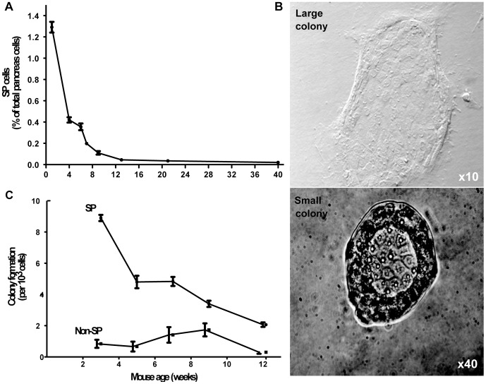
Figure 3. The proportion of SP cells and their colony forming potential decreases with age.
A) Pancreas cells from littermates aged 5 days to 40 weeks were stained with Hoechst 33342 dye to quantitate the SP by flow cytometry (n = 7–15 per group). B) Colonies formed by SP and non-SP cells isolated from pancreas of littermates aged 3 to 12 weeks were quantified (n = 7–10 per group). C) Two colony types were observed after 10 days of culture on Matrigel.

