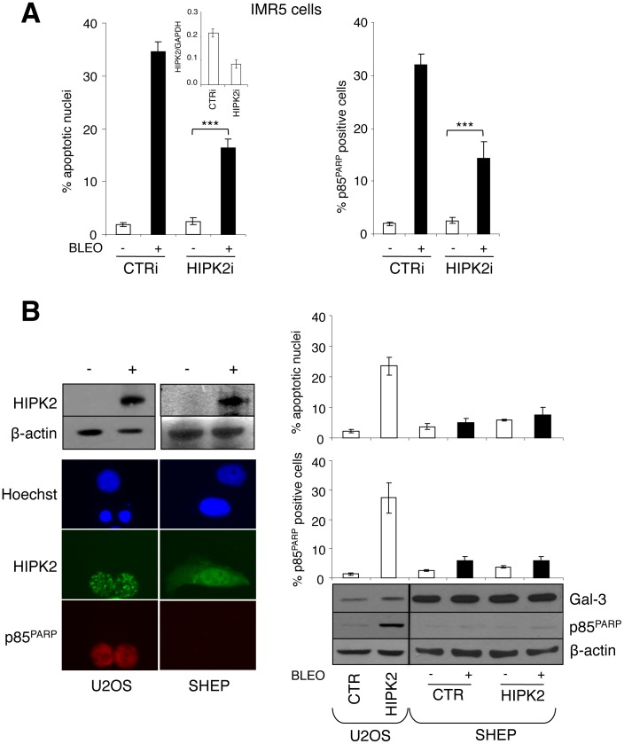
Figure 1. Exogenous HIPK2 expression is not sufficient to induce apoptosis or sensitizes MNSC cells to DNA damaging drugs. A.
, HIPK2 knock-down was achieved by transient transfection with sh-RNAi in MNA IMR-5 cells and was measured by Q-RT-PCR (inset). Analysis of bleomycin (5µg/ml) induced apoptosis is shown as percentage of apoptotic nuclei and cells positive for p85 cleaved fragment of the PARP protein (p85PARP). Significant differences in apoptosis fold induction were obtained between HIPK2i and CTRi transfected cells (***p<0.0001) B, Cell transfection with HIPK2 (+) but not empty vector (−) caused apoptosis in the U2OS osteosarcoma cells as indicated by the appearance of apoptotic nuclei and/or positive staining for p85PARP (left panel), but failed to induce apoptosis and to sensitize MNSC SHEP neuroblastoma cells to bleomycin treatment (percentage of the apoptotic cells are given in the graphs in the right panel). The immunoblot (lower right panel) shows the accumulation of the indicated proteins in HIPK2 transfected and/or bleomycin treated cell extracts.

