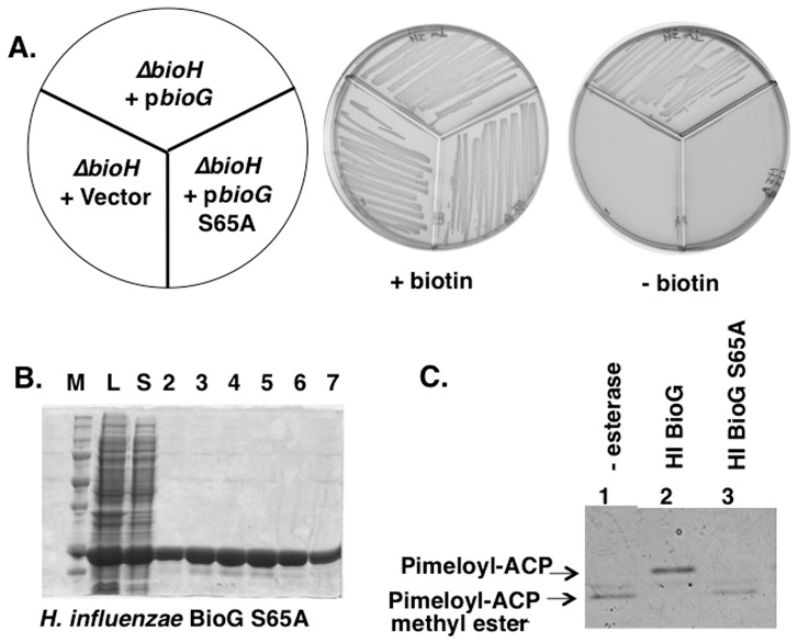
Figure 7. Loss of BioG function upon substitution of the putative active site serine with alanine.
Panel A. E. coli strain STL24 (ΔbioH) was transformed with plasmids encoding H. influenzae BioG S65A (right), wild type H. influenzae BioG domain (top) or the empty pET28b+ vector (left). The transformants were streaked onto M9 plates containing 0.2% glucose. Panel B Purification of the S65A BioG. Eluted fractions (10 µl) were analyzed by electrophoresis on a 10% SDS-polyacrylamide gel. The lysate and soluble fractions are shown in lanes L and S, respectively. Panel C. The H. influenzae BioG S65A protein was assayed for esterase activity as in Figure 6.

