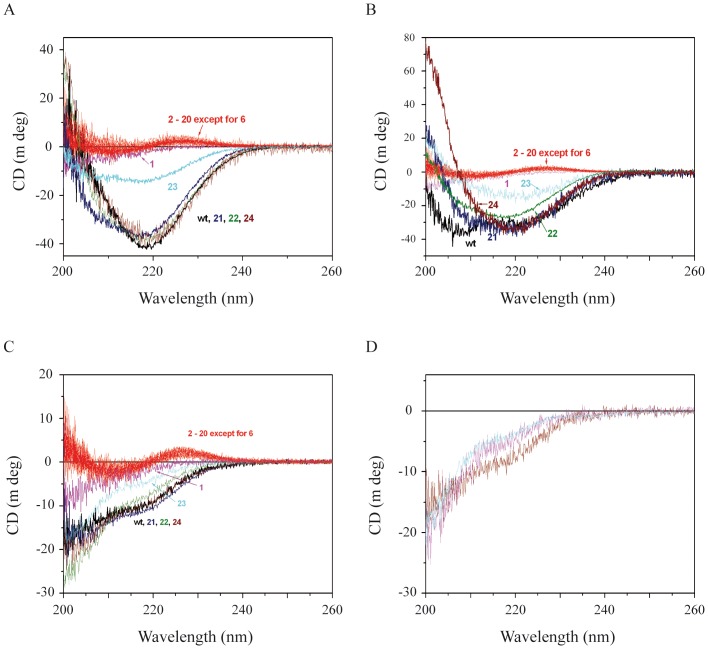
Figure 5. CD spectra of wt and alanine-substituted peptides.
CD spectra of 20 µM biotinylated wt and alanine-substituted NFL-TBS.40-63 peptides were measured in KF/phosphate buffer (Panel A), 2 mM SDS (Panel B) and pure water (Panel C). The spectra of wt peptide are in black. The spectra of peptides with a substitution by alanine at positions from 2 to 20 (except at position 6 where the original AA is alanine) are similar and represented by red lines. The spectra of wild-type (black), Tyr1Ala (magenta), Ser21Ala (dark blue), Ser22Ala (olive), Gly23Ala (light blue) and Ser24Ala (wine) are also presented. The number corresponds to the position of the replaced AA residue. CD spectra of 20 µM NFL-TBS.40-63 peptides phosphorylated at positions 17 (wine), 19 (magenta) and the two positions (cyan) were measured in KF/phosphate buffer (Panel D).

