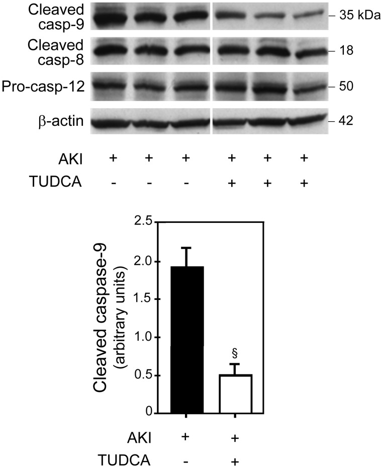
Figure 4. Apoptosis pathway analysis.
TUDCA treatment significantly blocked activation of caspase-9 following AKI as compared to vehicle treatment (rats 1, 2, and 3) (top panel). There was no difference in the activation of caspase-8 and caspase-12 between the TUDCA- and vehicle-treated rats. Densitometry analysis of cleaved caspase-9 normalized for β-actin (lower panel). When densitometry results for caspase-9 were compared between the TUDCA- and vehicle-treated groups, there was significantly less (p<0.01) activation of caspase-9 in the TUDCA group. Results are expressed as mean ± standard deviation of a least 3 different animals in each group. §p<0.01 from vehicle-injected controls.

