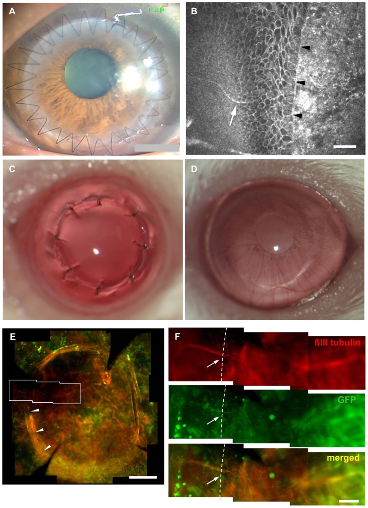
Figure 1. Peripheral nerve damage following corneal transplantation.
Corneal transplantation in humans invovles a 360 degree full thickness incision of the cornea (A), and in vivo confocal micorscopy reveals recipient nerve fibers (white arrow) extending to the donor-host junction (arrow heads)(B). None of the 6 patients exmined demonstrated signs of nerve regerenation within the donor at 3 months. A murine transplantation model was developed by transplanting 2 mm wild-type donors into P0-Cre/Floxed-EGFP hosts (C) Sutures were removed after 7 days to avoid excessive inflammation (D). Peripheral nerves can be observed extending to the donor host juntion (arrow heads in E) by positive βIII tubulin staining and GFP in a magnified view (F). Dotted line in (F) shows the border of the donor and recipient cornea. Scale bar = 50 µm in B, 500 µm in E and 100 µm in F.

