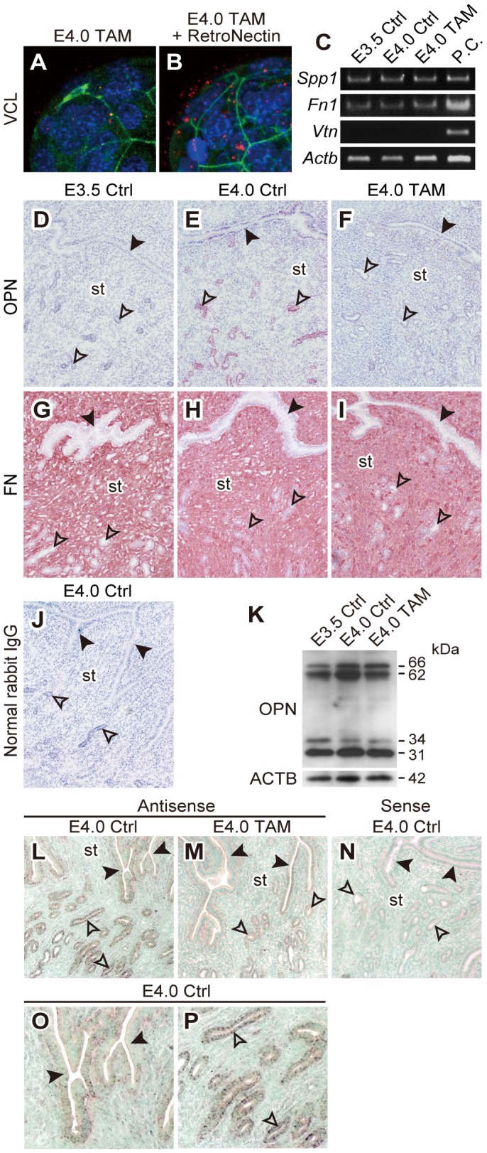
Figure 4. Endometrial glandular epithelial cells from the receptive uterus express osteopontin.

(A and B) Blastocysts collected from E4.0 TAM mice were incubated for 90 min with or without RetroNectin in DMEM/F12 medium. The effect of RetroNectin treatment on expression and distribution of VCL were then evaluated by immunofluorescence. Immunolocalization of VCL was visualized with Alexa 588 (red). F-actin and nuclei were stained with phalloidin-Atto 488 (green) and DAPI (blue), respectively. Z-stack projection images of confocal laser-scanning microscopy are presented. Multiple samples were analyzed on at least three separate occasions. (C) Expression of Spp1, Fn1 and Vtn mRNAs in E3.5 Ctrl, E4.0 Ctrl and E4.0 TAM uteri were evaluated by RT-PCR. Total RNAs from three uteri for each group were analyzed. P.C., positive control. (D–J) Immunohistochemistry for OPN (D–F) and FN (G–I) was performed on uterine tissues from E3.5 Ctrl, E4.0 Ctrl or E4.0 TAM mice. Normal rabbit IgG served as a negative control (J). Uterine tissues from at least three animals for each group were analyzed. Representative results are presented. Black arrowheads: luminal epithelium, open arrowheads: glandular epithelium, st: stroma. (K) Western blotting for OPN in protein extracts from E3.5 Ctrl, E4.0 Ctrl and E4.0 TAM uteri. Uterine tissues from at least three animals for each group were analyzed. Representative blot is presented. β-actin was used as loading control. Four species of immunoreactive OPN were found at molecular weights of approximately 66 kDa, 62 kDa, 34 kDa and 31 kDa. (L–P) In situ hybridization for Spp1 transcripts on E4.0 uterine tissues. Antisense (L and M) and sense (N) probes for Spp1 mRNA were hybridized on uterine tissues from E4.0 Ctrl (L and N) and E4.0 TAM (M) mice. (O and P) Higher magnification images of L highlighting luminal epithelia (O) and glandular epithelia (P) of E4.0 Ctrl uterus. Note that the expression of Spp1 mRNA on E4.0 uterus (L) was more prominent in glandular epithelia (P) than that in luminal epithelia (O) and disrupted by TAM treatment (M). Sense probe did not show any signals on E4.0 uterus (N). Uterine tissues from at least three animals for each group were analyzed. Representative results are presented. Tissue sections were counter-stained with methyl green. Black arrowheads: luminal epithelium, open arrowheads: glandular epithelium, st: stroma.
