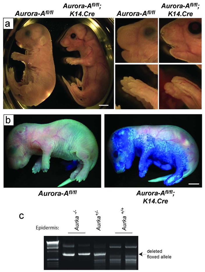
Figure 1. Skin abnormalities in mice deficient for epithelial Aurora-A.

(a) Left panels show gross phenotypic presentation of Aurora-Afl/fl; K14.Cre mice at E18.5. These mice show translucent skin compared to WT controls. Right panels show a close up of the face and paws. Note the improperly formed whiskers, nares, eyelids and nails. (b) Toluidine blue dye penetration assays revealed a defect in barrier function in Aurora-Afl/fl; K14.Cre E18.5 mice. (c) Tails from newborn mice were digested and PCR performed to detect Cre-mediated recombination of the floxed Aurora-A allele. Bars= 2.5 mm.
