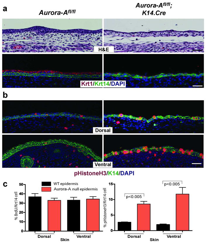
Figure 3. Delayed stratification in Aurora-A−/− epidermis.

(a) Epidermis between E13–14 was examined by histology and marker analysis. Unlike WT mice that forms two layers at E13.5, Aurora-A−/− epidermis remains primarily a single layer. Consistently, few cells expressed the subrabrasal marker Krt1 in the uppermost layer of the skin. A similar pattern was observed on ventral skin (not shown). (b) Immunofluorescent detection of phopho Histone H3 (Ser10) in E13.5 skin. Note the increased number of positive cells found in both dorsal and ventral skins of Aurora-Afl/fl; K14.Cre mice. (c) Quantitation of BrdU incorporation and detection of mitotic cells per Krt14 positive keratinocytes. Data shown is the average counts per three embryos ± standard deviation. P values are from a t-test. Bar = 25 μm
