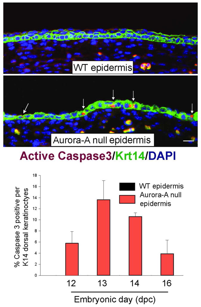
Figure 4. Increased apoptosis in Aurora-A deficient epidermis.

Top panel shows the immunodetection of active Caspase 3 in WT and Aurora-A−/− epidermis at E13.5. Bar= 25 μm. Bottom panel shows the quantitation of apoptosis found in developing skin of Aurora-Afl/fl; K14.Cre or WT embryos (n=2 embryos per genotype and timepoint). Columns represent average ± standard deviation. Note the absence of apoptotic cells in WT skin at any embryonic stage.
