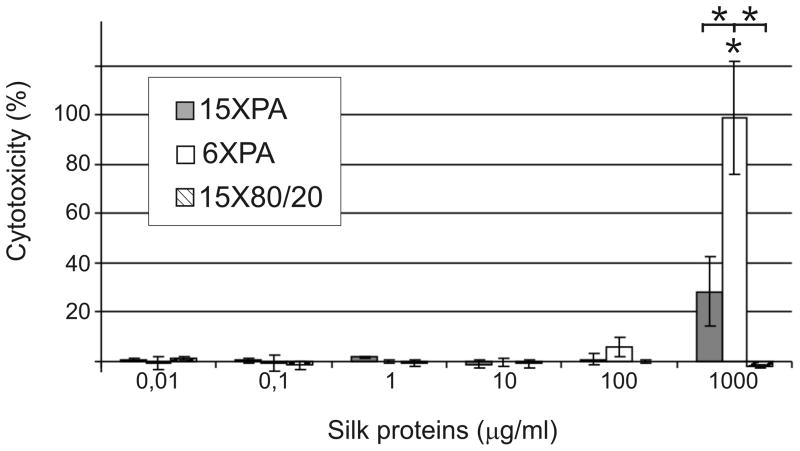
Figure 2.
Cytotoxicity of spider silk proteins evaluated by LDH released into culture medium. NIH 3T3 cells were maintained in medium supplemented with different concentrations of spider silk proteins for 24h. The amount of LDH released was measured by spectrophotometric analysis and cytotoxicity was calculated as described in the Materials and Methods. Results are expressed as the means of three independent experiments and error bars show the standard deviation.

