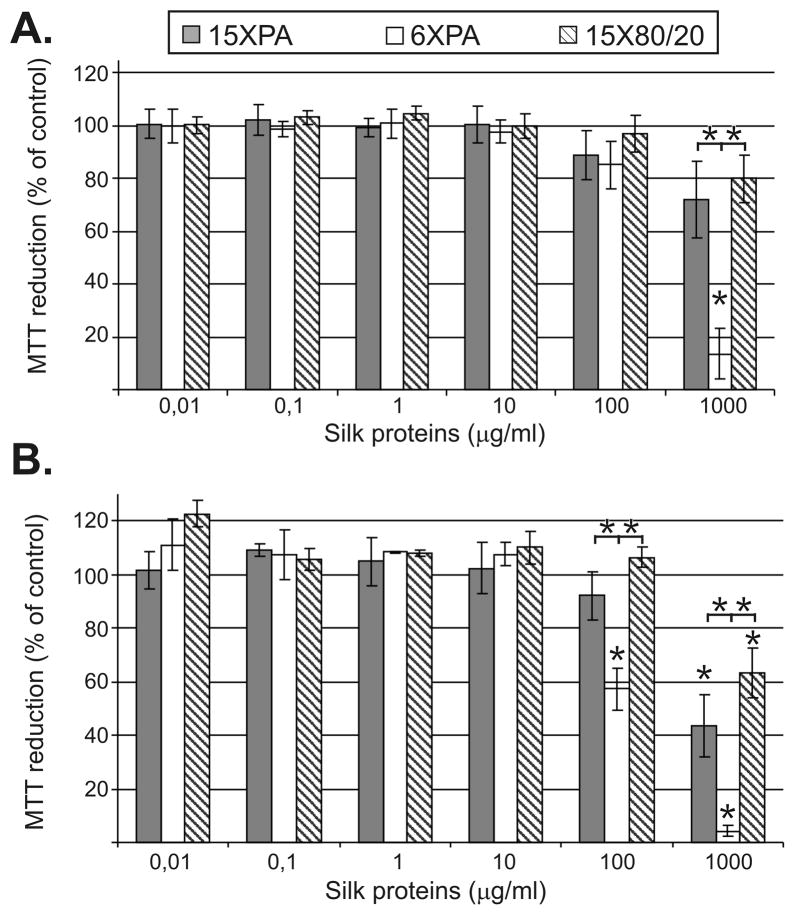
Figure 3.
Mitochondrial activity assessed using MTT. NIH 3T3 cells were cultured in the presence of the spider silk proteins for 24h (A) and 48h (B). The % of the MTT reduction was calculated as described in the Materials and Methods. Results are expressed as means of three independent experiments and error bars show the standard deviation.

