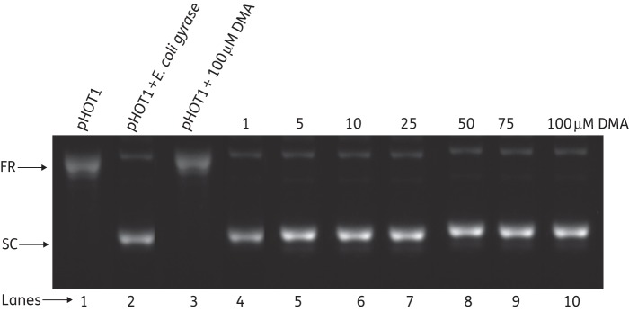
Figure 7.
Analysis of supercoiling of relaxed pHOT1 by E. coli gyrase in the presence of DMA. Lane 1, relaxed DNA as control; lane 2, supercoiling of relaxed DNA by E. coli DNA gyrase; lane 3; relaxed DNA in the presence of 100 μM DMA; lanes 4–10, supercoiled DNA in the presence of 1, 5, 10, 25, 50, 75 and 100 μM DMA, respectively. FR, fully relaxed; SC, supercoiled.

