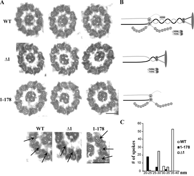
Figure 6.
Distinct stubby spokes in the axonemes from the Δ1 and 1–178 mutants. (A) The representative transmission electron microscopic images of cross-sectioned axonemes from the WT, Δ1, and 1–178 strains. The bottom panel gives an enlarged view of the axoneme cross sections. The arrows highlight representative RSs in each strain. The enlarged spoke head is present in the RSs of WT and Δ1 axonemes. Bars, 100 nm. (B) Schematic pictures depicting the RSs in each strain. (C) The length distributions of RSs in cross-sectioned axonemes. The RSs with an identifiable morphology were measured from 13 WT sections, 24 Δ1 sections, and 22 1–178 sections. RSs were separated based on the spoke length, and the number in each group was plotted into a distribution histogram.

